

Table of Contents
- 1. Enhanced Diagnostic Accuracy With 3D Cone Beam Imaging
- 2. Complete Treatment Planning Before Your First Appointment
- 3. Precise Implant Placement and Prosthodontics Success
- 4. Early Detection of Hidden Decay and Disease
- 5. Reduced Radiation Exposure Compared to Traditional X-rays
- 6. Real-Time Visualization for Patient Confidence and Consent
- 7. Streamlined Cosmetic Smile Design and Makeover Planning
- Frequently Asked Questions (FAQ)
1. Enhanced Diagnostic Accuracy With 3D Cone Beam Imaging
Two-dimensional X-rays have been the dental standard for decades, but they come with a fundamental limitation: they flatten complex three-dimensional anatomy onto a flat image. Imagine trying to understand the layout of your entire home by looking at only its shadow. You’d miss crucial details about room placement, depth, and spatial relationships.
3D cone beam imaging eliminates this guesswork. Our X-MIND PRIME system captures over 600 individual images from multiple angles, then reconstructs them into a complete volumetric view of your teeth, jaw, and bone structure. This means we can:
- Identify bone density and quality before implant placement
- Detect conditions like impacted teeth or jaw pathology that 2D imaging would overlook
- Measure exact distances between anatomical structures with millimeter precision
- Spot calcifications, cysts, or abnormalities that could otherwise go unnoticed
When we can see exactly what we’re working with, treatment plans become far more predictable. You avoid surprises during procedures, and we deliver results that are reproducible and reliable. This accuracy is especially critical when planning complex restorative work or emergency interventions where precision directly impacts your long-term outcome.
Your next move: Schedule a consultation so we can capture a baseline 3D scan. This imaging becomes your diagnostic foundation for all future dental decisions.
2. Complete Treatment Planning Before Your First Appointment
One of the most stressful aspects of dental treatment is the unknown. You walk in, sit down, and then discover what you actually need. With 3D imaging, we flip that script entirely.
Before you ever sit in the treatment chair, we’ve already mapped out your entire clinical picture. Using our digital imaging data, we can design your complete treatment pathway, identify any risk factors, and prepare all necessary materials and instruments in advance. This preparation means:
- Your appointment runs on schedule without unexpected delays
- We explain exactly what to expect because we’ve already visualized every step
- We reduce chair time and associated discomfort
- We eliminate the anxiety that comes from clinical surprises
For patients requiring smile makeovers or complex restorative cases, this level of pre-planning is transformative. You can see renderings of your proposed smile design and understand precisely how we’ll rebuild your dentition. You’re not walking in blind; you’re walking in informed and confident about what comes next.
Your next move: Request our pre-appointment consultation where we’ll review your 3D scans and walk you through the entire treatment timeline and cost breakdown.
3. Precise Implant Placement and Prosthodontics Success
Dental implants are an investment in your future. A misplaced implant can compromise esthetics, function, and longevity. A perfectly placed implant can last decades and feel completely natural.
3D imaging is the difference between those two outcomes. Before implant surgery, we use cone beam CT to analyze:
- Bone volume and density at the intended implant site
- The position of vital structures like the inferior alveolar nerve and sinus
- Optimal implant diameter, length, and angulation
- Bone width, height, and quality to determine if grafting is necessary
With this intelligence, our surgical planning becomes a precise mathematical exercise rather than a guessing game. We can even perform virtual implant placement to verify that the final restoration will emerge at the ideal angle and position for maximum esthetic and functional success.
In prosthodontics, where we’re restoring or replacing multiple teeth, 3D imaging allows us to create customized restorations that integrate seamlessly with your existing anatomy. The result is implants and bridges that look natural, function like biological teeth, and deliver the confidence that comes from knowing your restoration was engineered for success.
Your next move: If you’re considering implants, bring your initial 3D scan to discuss placement options and bone grafting needs before committing to surgery.
4. Early Detection of Hidden Decay and Disease
Decay doesn’t always announce itself. Small cavities hide between teeth or beneath existing restorations where a traditional X-ray might miss them entirely. By the time symptoms appear, the damage has often progressed significantly.
Our SOPROLIFE decay detection technology works alongside 3D imaging to catch problems at their earliest, most treatable stage. We can identify:
- Interproximal cavities (decay between teeth) before they become visible
- Recurrent decay under old fillings or crowns
- Subtle bone loss associated with gum disease
- Periapical pathology around tooth roots
Early detection means smaller, more conservative treatments. Instead of a large filling or root canal, we catch the issue when a small intervention stops it cold. This approach preserves more of your natural tooth structure and saves you time, money, and future complications.
For patients with a history of decay or periodontal concerns, this proactive surveillance becomes part of your long-term oral health strategy. You’re not just treating problems as they appear; we’re staying ahead of them.
Your next move: Schedule regular 3D scans annually if you have a history of cavities or gum disease to catch problems before they require extensive treatment.
5. Reduced Radiation Exposure Compared to Traditional X-rays
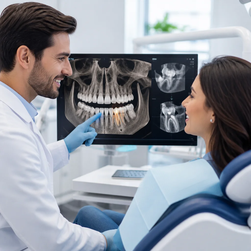
A common concern about advanced imaging is radiation safety. Ironically, 3D cone beam imaging delivers a complete diagnostic picture while exposing you to less radiation than multiple traditional X-rays.
Here’s why: A full-mouth series of conventional X-rays might include 14 to 18 individual films, each requiring positioning and re-exposure. In contrast, our cone beam CT captures everything in a single, quick rotation lasting just 10 to 20 seconds. The result is lower cumulative dose and faster imaging.
We also practice the principle of “ALARA” (As Low As Reasonably Achievable) by:
- Using lead aprons and thyroid collars to shield sensitive tissues
- Selecting imaging protocols appropriate to your specific clinical situation
- Avoiding unnecessary scans by optimizing our imaging schedule
- Keeping detailed records to prevent redundant exposures
Advanced imaging should never compromise your safety. At Smile Expert Design Center, we’ve chosen technology that prioritizes both diagnostic excellence and radiation safety because your long-term wellbeing matters more than any single scan.
Your next move: Ask us about our radiation safety protocols during your next visit, and request that we maintain your imaging records to prevent duplicate scans.
6. Real-Time Visualization for Patient Confidence and Consent
Dental anxiety often stems from a lack of control and understanding. You’re in the chair, someone’s working in your mouth, and you have no idea what they’re seeing or why they’re doing what they’re doing. That’s unsettling.
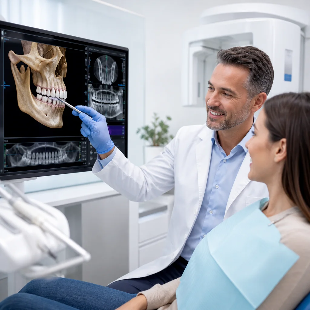
3D imaging changes that dynamic completely. We can show you exactly what we see on a screen right in front of you. You can rotate the image, zoom in on problem areas, and understand the clinical rationale for our recommendations. This transparency builds trust because:
- You’re no longer relying on our word alone; you can see the evidence yourself
- Treatment becomes collaborative rather than directive
- You understand the consequences of treatment versus no treatment
- You feel genuinely involved in decisions about your own mouth
When we’re planning a cosmetic procedure or smile makeover, visualization becomes even more powerful. You can see the 3D renderings of your proposed smile and provide feedback before we place a single crown or aligner. This iterative design process ensures your final result aligns perfectly with your aesthetic vision and expectations.
Your next move: During your next appointment, ask to review your 3D scan images on our visualization screen so you can fully understand your treatment options.
7. Streamlined Cosmetic Smile Design and Makeover Planning
Your smile is often the first thing people notice about you. A smile makeover should enhance your natural beauty and boost your confidence, not create something that looks “done” or artificial.
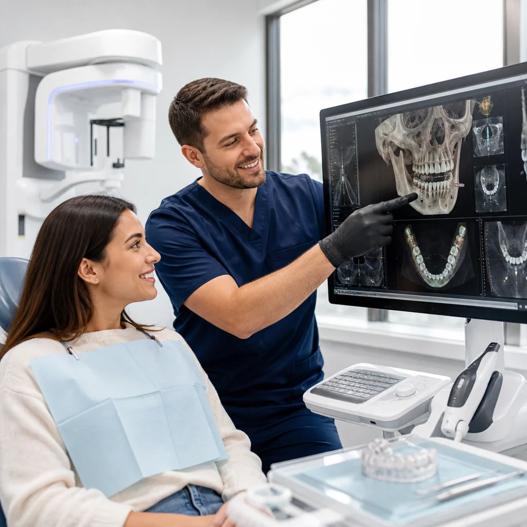
With our precision digital smile design approach, 3D imaging enables us to:
- Analyze your facial symmetry, lip line, and dental midline in three dimensions
- Preview multiple smile design options and show you how each looks on your face
- Design restorations that harmonize with your bone structure and proportions
- Plan shade, shape, and contour to match your personal aesthetic goals
The combination of 3D imaging and digital smile previews means you know exactly what your new smile will look like before treatment begins. No surprises. No regrets. Just a smile that’s authentically, beautifully yours and dramatically improved from where you started.
For comprehensive smile makeovers involving veneers, crowns, or clear aligner orthodontics, this level of digital planning transforms the entire experience. You walk out of our office not just with a better smile, but with the confidence that comes from seeing your transformation in advance and approving every detail.
Your next move: Book a smile design consultation where we’ll create multiple 3D renderings of your potential smile and discuss which design resonates most with your vision.
For further reading: Digital smile previews.
Frequently Asked Questions (FAQ)
How does your 3D digital imaging technology improve my treatment outcomes?
We use X-MIND PRIME 3D digital imaging to capture comprehensive views of your teeth, bone structure, and facial anatomy in a single scan. This allows us to identify problems that traditional 2D X-rays miss and plan your treatment with precision, whether you’re getting implants, cosmetic work, or restorative care. Our detailed imaging means we can execute your treatment exactly as planned, delivering results you can trust.
What’s the difference between your 3D imaging and standard dental X-rays?
Our cone beam imaging provides a complete three-dimensional picture of your mouth, while traditional X-rays show only flat, two-dimensional views. We expose you to significantly less radiation with our advanced system, and we can detect decay, bone loss, and other issues much earlier. This technology lets us catch problems before they become serious and more expensive to treat.
Can I see my smile design before we start cosmetic or restorative treatment?
Yes. We use our 3D imaging system to show you exactly how your new smile will look before any work begins. This real-time visualization helps us collaborate with you on the design, answer your questions, and ensure you’re completely confident in the plan we’ve created together.