

Table of Contents
- Why Traditional Bite Assessment Falls Short for Aligner Therapy
- The Challenge of Predicting Complex Bite Corrections
- How Our X-MIND PRIME 3D Imaging System Works
- Capturing Complete Anatomical Data for Precision Planning
- Creating Detailed Digital Treatment Maps Before Aligner Fabrication
- Reducing Complications Through Predictive Analysis
- The Patient Experience: From Scan to Custom Aligners
- Achieving Predictable Results for Complex Cases
- Why Visualization Matters in Aligner Success
- Our Commitment to Evidence-Based Smile Transformation
- Frequently Asked Questions (FAQ)
Why Traditional Bite Assessment Falls Short for Aligner Therapy
Clear aligner therapy has revolutionized orthodontics by offering discreet, comfortable tooth straightening without metal brackets. Yet the success of any aligner case depends entirely on precise treatment planning before a single tray is fabricated. At Smile Expert Design Center, we’ve moved beyond conventional bite assessment to leverage advanced 3D imaging technology that fundamentally changes how we predict, plan, and deliver aligner results. Our X-MIND PRIME 3D imaging system gives us the complete picture needed to address complex bite corrections with confidence and accuracy.
Two-dimensional X-rays and plaster molds have served dentistry for decades, but they capture only a fraction of the information needed for modern aligner planning. Traditional bite records show tooth position from one angle and don’t reveal how the jaw joint functions, how bone structure supports tooth movement, or where real obstacles to alignment exist in three dimensions.
When we rely solely on flat images, we miss critical details:
- Bone density variations that affect how quickly teeth move
- Asymmetries in the jaw that a 2D photo cannot show
- The true vertical relationship between upper and lower teeth
- Root positioning beneath the gumline
- Soft tissue contours that impact the final aesthetic result
Patients sometimes return after months of aligner wear only to discover their bite hasn’t improved as expected, or that certain teeth haven’t moved as planned. This happens not because aligners fail, but because the initial planning lacked the depth of information needed to create an accurate roadmap. We’ve seen too many cases where traditional bite assessment simply couldn’t predict the challenges ahead.
The Challenge of Predicting Complex Bite Corrections
Bite correction is far more intricate than simply moving teeth closer together. Every tooth has a root beneath the surface, and every root sits within bone that varies in density and height from one area of the mouth to another. When we attempt to correct an overbite, underbite, or crossbite with incomplete information, we’re essentially navigating in dim light.
Complex cases require us to anticipate:
- How much bone remodeling will occur as teeth shift positions
- Whether impacted or severely angled teeth need preliminary intervention
- If the jaw joint can accommodate the new bite position comfortably
- Whether asymmetries in bone structure will limit certain movements
- Which teeth need to move first to create space for others
Consider a patient with a moderate underbite and crowded lower teeth. Traditional planning might suggest a standard aligner sequence, but three-dimensional imaging might reveal that one side of the lower jaw is narrower than the other, which means our tooth movement strategy needs to be asymmetrical. Without that insight, the treatment could stall halfway through or leave the bite uncorrected despite months of wear.
How Our X-MIND PRIME 3D Imaging System Works
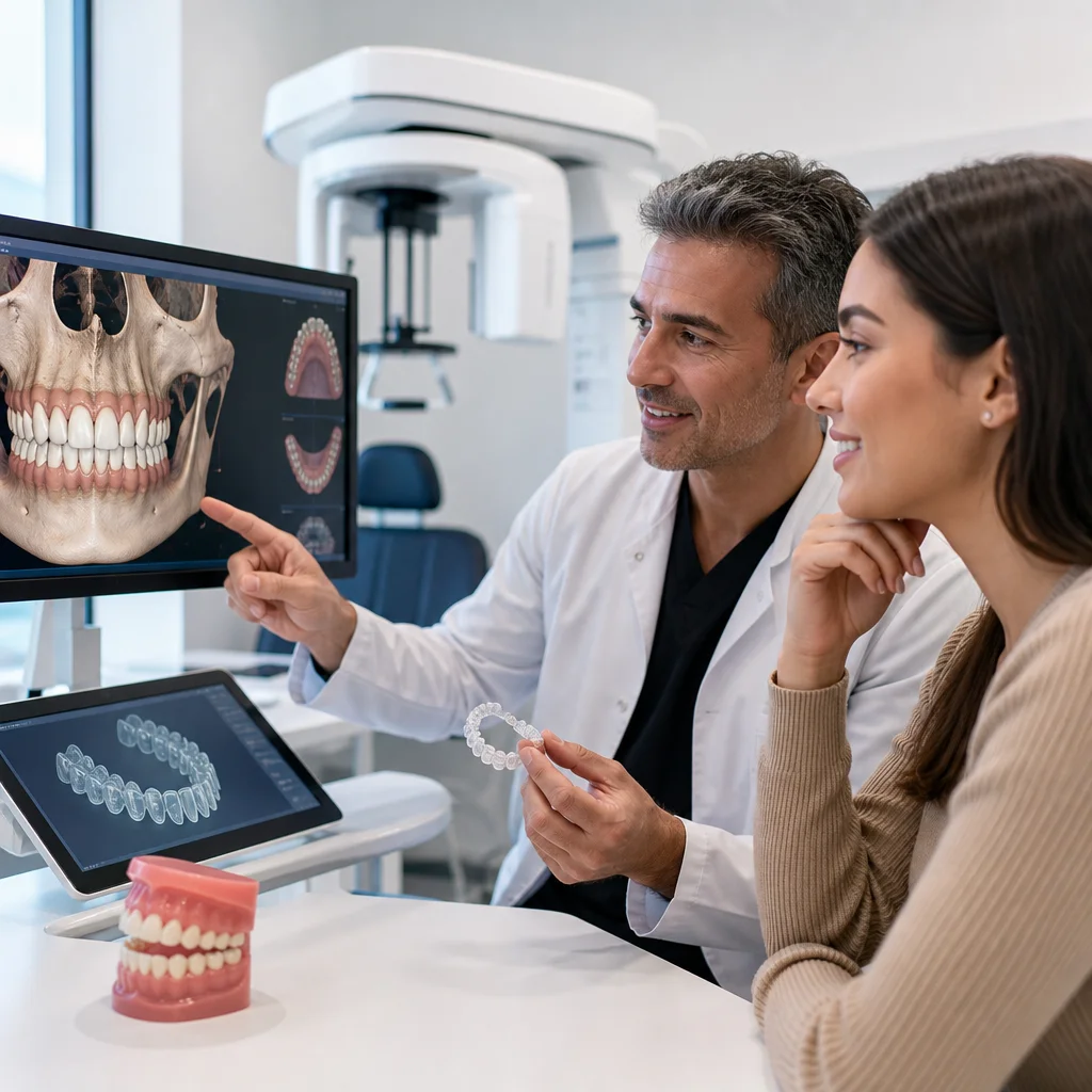
Our X-MIND PRIME cone beam imaging technology captures a complete volumetric dataset of your entire dentofacial structure in a single scan. Unlike conventional flat-panel X-rays, this system rotates around your head and collects hundreds of images that are reconstructed into precise three-dimensional models.
The scan takes roughly 12 seconds and delivers:
- Full jaw and skull geometry with submillimeter accuracy
- Complete tooth root anatomy, including length, curvature, and orientation
- Bone volume and density mapping across the entire treatment area
- Soft tissue contours that influence smile aesthetics
- Temporomandibular joint position and structure
- Airway space and sinus anatomy for safety assessment
The radiation dose is significantly lower than traditional comprehensive X-ray series, which means we gather vastly more useful information with less exposure. We can even identify issues that would require separate imaging with older technology, reducing your need for repeat scans.
Capturing Complete Anatomical Data for Precision Planning
Once we have your three-dimensional scan, specialized software allows us to measure and analyze every aspect of your bite anatomy. We’re not interpreting shadows on film; we’re examining actual digital models that can be rotated, sectioned, and measured from any angle.
Our team examines:
- Exact distances between upper and lower teeth in vertical, horizontal, and anteroposterior dimensions
- Root length and position relative to bone boundaries
- Bone height and density in areas where tooth movement will occur
- Asymmetries between the left and right sides of your jaw
- The relationship between your jaw and skull base
- Gum tissue thickness and potential areas of recession risk
This level of detail allows us to predict tooth movement with far greater accuracy than traditional methods. If severe bone resorption is visible in one area, we know to move teeth more carefully there. If a tooth root is very curved or angled, we know to apply gentler forces over a longer timeframe. Every critical variable is visible and measurable before treatment begins.
Creating Detailed Digital Treatment Maps Before Aligner Fabrication
With complete anatomical data in hand, we use specialized treatment planning software to create a step-by-step movement sequence customized to your anatomy. This isn’t guesswork; it’s a precise digital blueprint built from your actual measurements.
We determine:
- The sequence in which each tooth should move
- How much tooth movement is possible in each direction
- Where intermediate checkpoints are needed to verify progress
- Which teeth might require additional support or springs
- The optimal thickness and force level for each aligner stage
- Whether your case requires interproximal reduction (slight space between teeth) to accommodate crowding
- Realistic timelines based on your individual bone response
The digital map shows us before fabrication begins whether your case is straightforward or requires additional planning steps. Some patients benefit from selective tooth extractions or preliminary treatment before aligner therapy begins. Others need temporary anchoring devices or modified aligner designs. We identify these factors upfront, not after three months of ineffective tooth movement.
Reducing Complications Through Predictive Analysis
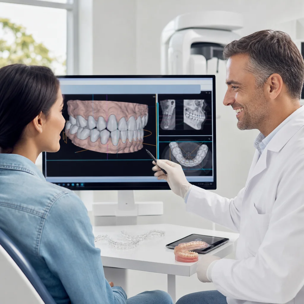
One of the most valuable aspects of advanced 3D planning is its ability to identify potential problems before they develop. We can virtually simulate tooth movement and watch for areas of concern:
- Roots that might move outside the bone boundaries (a serious complication called root resorption)
- Teeth that might collide with other teeth or bone
- Areas where gum tissue might recede if teeth move too aggressively
- Asymmetric movement patterns that could skew your midline
By predicting these challenges, we modify the treatment plan to avoid them. Perhaps we move one area more slowly. Perhaps we need two additional aligner phases. Perhaps a tooth needs to move in a different sequence than initially planned. These adjustments happen in our digital planning phase, not during your treatment, which means fewer surprises and faster overall results.
Patients appreciate knowing that we’ve already thought through potential obstacles and built safeguards into their treatment design. It’s the difference between a dentist responding to problems as they arise and one who prevents them from occurring in the first place.
The Patient Experience: From Scan to Custom Aligners
Your aligner journey begins with a comfortable 12-second cone beam scan in our office. You stand still while the machine rotates around your head, and there’s no pain or discomfort. We don’t need bite impressions or temporary models because the 3D data is far more detailed and accurate.
Within days, our treatment team reviews your complete digital anatomy and creates your customized plan. We then show you a digital smile design preview that illustrates your projected results. You’ll see exactly how your teeth will move, what your new bite will look like, and how many aligner stages your treatment will require.
Once you approve the plan, your custom aligners are fabricated using 3D models generated directly from your imaging data. Every aligner in your series is designed specifically for your anatomy, not based on generic movement templates. This personalization is why aligner therapy succeeds at our practice where other approaches might stall.
Achieving Predictable Results for Complex Cases
Complex cases that once required metal braces or surgical intervention are now successfully treated with aligners when planned correctly. A patient with a moderate underbite, crowded teeth, and asymmetric jaw growth can receive a comprehensive aligner plan that accounts for all these variables simultaneously.
We’ve successfully treated patients with:
- Severe crowding requiring strategic tooth movement sequencing
- Significant bite corrections (overbites, underbites, crossbites)
- Asymmetric jaw development
- Prior orthodontic relapse
- Missing or congenitally small teeth
- Compromised bone levels from prior periodontal disease
Each of these cases demands precision planning that only 3D imaging and advanced analysis can provide. The results are predictable because we’ve built them into the plan from the beginning.
Why Visualization Matters in Aligner Success
Patients invest months and significant resources in aligner therapy. They want to understand their treatment fully and see the path to their new smile. Advanced 3D imaging allows us to show you exactly what we’re doing and why.
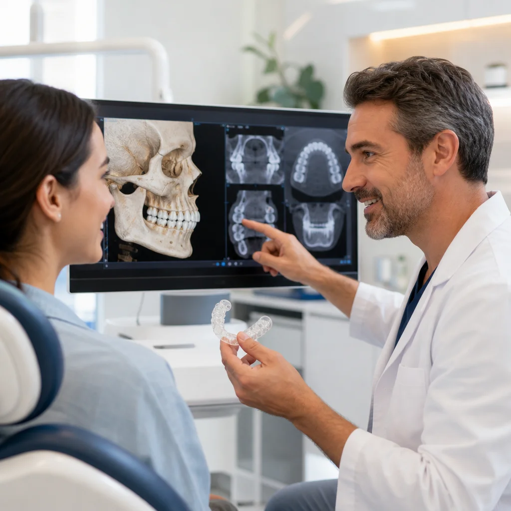
Visualization serves several purposes:
- It builds confidence by showing that your unique anatomy has been thoroughly analyzed
- It clarifies why your treatment timeline is appropriate for your specific case
- It demonstrates our clinical sophistication and attention to detail
- It helps you stay motivated when you can track progress against the predicted plan
- It creates accountability for both our practice and your compliance with wear instructions
When patients understand their treatment at this level, they’re more invested in success and more likely to wear aligners consistently. They see how their teeth are moving precisely as predicted, which reinforces that the plan was sound and the investment worthwhile.
Our Commitment to Evidence-Based Smile Transformation
At Smile Expert Design Center, Dr. Jean-Jacques Edderai and our team are committed to delivering exceptional results through technology and clinical expertise. Advanced 3D imaging for aligner treatment planning represents the standard of care we’ve chosen to provide because your smile transformation deserves precision, not approximation.
We combine our X-MIND PRIME imaging system with precision digital smile design methodology to ensure your new smile improves not just alignment but also aesthetics, function, and long-term oral health. Every aligner you wear is based on detailed knowledge of your unique anatomy.
If you’re considering clear aligner therapy and want treatment planned with complete anatomical precision, we’d welcome the opportunity to scan your smile and discuss your options. Our North Miami Beach practice serves patients throughout Miami, North Miami Beach, and Miami Beach with comprehensive family dentistry, advanced cosmetic care, and the technology to make confident smile transformations real.
Contact us to schedule your 3D imaging consultation and see exactly how we would plan your personalized aligner treatment.
Frequently Asked Questions (FAQ)
How does our X-MIND PRIME 3D imaging improve aligner treatment compared to traditional methods?
Our X-MIND PRIME system captures your complete dental and jaw anatomy in three dimensions, allowing us to see bite patterns and tooth positioning that standard 2D X-rays simply cannot reveal. We use this detailed mapping to predict exactly how your teeth will move throughout your aligner treatment, which means we can anticipate and prevent complications before they occur. This precision planning helps us achieve results that are more predictable and often faster than conventional approaches.
What happens during the 3D scanning process at our practice?
We position you in our X-MIND PRIME imaging unit for a quick, comfortable scan that takes just seconds and exposes you to minimal radiation. Our system generates a complete digital model of your teeth, jaw structure, and bite relationship, which we then analyze to create your personalized treatment plan. You’ll see your own scan results and digital smile preview, so you understand exactly what we’re working toward before your custom aligners are fabricated.
Can our advanced imaging handle complex bite problems that other practices might turn away?
Yes, we specifically invested in X-MIND PRIME technology because it lets us successfully treat cases with significant bite corrections, crowding, or spacing issues that require precise predictive analysis. We analyze your unique anatomy through our 3D data to determine whether aligners are right for you or if we need to recommend alternative restorative solutions. This evidence-based approach means you get honest answers about what’s achievable for your particular situation.